
A Student Sonographer’s Cheat Sheet to a Successful Abdominal Ultrasound Exam

Do you have an abdominal ultrasound exam coming up and feeling a bit nervous about it? As a sonographer, I’ve got you covered. In this cheat sheet, we’ll go through everything you need to know, from what to expect during the exam to how to prepare yourself for it. We will also cover some tips and tricks to ace your abdominal ultrasound exam as a both a patient and a student sonographer. Follow along as we go into detail about how we scan for specific organs, to the difficulties faced when patients have different body types. We also discuss the common pathologies seen on abdominal ultrasound examinations and the reasons why people usually have them performed. By the end of this article, you’ll have all the knowledge you need to be prepared and confident for your exam.
What is an abdominal ultrasound exam?

An abdominal ultrasound exam is a radiological procedure used to examine the organs and structures in the abdominal cavity. It is performed similarly to when you have your fetus scanned in pregnancy. The difference being, at that exam we focus on things like femur, and humerus bones, baby’s circulatory system, vein, and arteries, and much much more …ONLY to do with your baby! This time, the exam is solely focused on you. This non-invasive imaging test is conducted by a diagnostic sonographer and provides detailed images of the organs and structures in the abdominal area, such as the liver and gallbladder. The ultrasound examination uses sound waves to create images of the internal organs, including blood flow through the abdominal organs and vessels. The images obtained during the exam help healthcare providers diagnose and treat diseases and conditions affecting the abdominal organs and structures. With proper preparation and understanding, patients can have a successful abdominal ultrasound exam.
Benefits of an abdominal ultrasound exam
An abdominal ultrasound is a useful diagnostic tool for assessing the condition of abdominal organs and diagnosing diseases. It is a non-invasive procedure that uses sound waves to create images of the organs in the abdomen. By providing an accurate picture of the internal organs, an abdominal ultrasound can detect tumours, cysts, or other abnormalities that would not otherwise be visible on an X-ray. The results of an abdominal ultrasound can also help guide the treatment plan for a patient, making it a valuable tool in the diagnostic process.
Preparation for an abdominal ultrasound exam

Preparation is key for a successful abdominal ultrasound exam. Depending on the type of exam, it may be necessary to fast for several hours before the appointment. The reason for this is to ensure that the gallbladder will be well imaged. When you eat, the gallbladder physiologically contracts, making it difficult to visualize, therefore difficult to rule out any pathology within it. It is for this reason that if you have eaten, your test may have to be rescheduled. It is also recommended that you wear comfortable clothing and remove any jewelry or metal objects that may interfere with the exam.
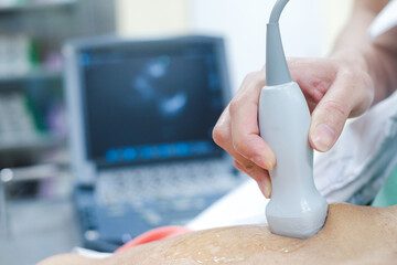
During the exam, a sonographer may will use a handheld device to capture images of the abdomen. These images can be used for diagnostic purposes to identify potential health issues. Before the exam, it is important to inform the clinician of any allergies or medications that you are currently taking.
After the exam is completed, the clinician may or may not discuss the results with the patient. With proper preparation and communication between the patient and clinician , an abdominal ultrasound exam can be a valuable tool for assessing and maintaining a patient’s health.
How to ace an abdominal ultrasound exam as a student sonographer
Preparing for abdominal ultrasound exams may require time and effort, but it is worth it in the long run. Understanding the anatomy of the patient and physics of the ultrasound machine is key to identifying any pathologies during the exam accurately. It’s essential to have a complete understanding of the specific protocols required for the test.
It’s always advisable to utilize the knowledge of an experienced sonographer who can provide tips and guidance to help you succeed during the exam. Furthermore, building a good rapport with patients can also help to improve their comfort level during the exam, making it easier for both you and the patient. With the right preparation, knowledge, and guidance, you can undoubtedly ace any abdominal ultrasound exam that comes your way.
Scanning the Liver Cheat Sheet:
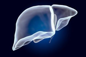
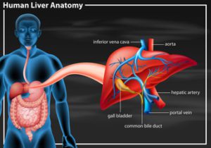
When it comes to acing an abdominal ultrasound exam, understanding how to properly scan the liver is crucial. Ultrasound can be used to scan and examine the liver for radiological emergency services, as well as aiding general practitioners in diagnosing medical conditions. This non-invasive method can identify any lesions on the liver, such as cysts, tumours, or abscesses, without the need for surgery. Additionally, ultrasound can detect any changes in the liver over time, making it an essential tool in monitoring liver health. Properly scanning the liver during an abdominal ultrasound exam is essential for accurate diagnosis and treatment planning. Both lobes of the liver should be scanned transversely and sagittally to include:
-Longitudinal (Sagittal). Left lobe. Caudate Lobe. IVC. Porta hepatis.
-Transverse . Left Lobe. Left hepatic vein. Left portal vein. Right portal vein.
-Middle and Right hepatic vein.
-Demonstrate hepatopetal flow in portal vein.
-Demonstrate hepatic vein flow.
What to look for: Check for diseases such as liver cancer, hepatitis , or cirrhosis. Lesions such as tumours, abscesses, or cysts of the liver or any trauma to the liver.
Note: Cirrhotic livers will look lumpy and shrunken as they will have advanced fibrosis compared to fatty livers.
Normal size of liver: 7-10.5 cm approximately.
Hepatic doppler ultrasound is also a standard part of the examination. It is used to assess the patency of the hepatic vasculature and the direction and velocity of the blood flow.
Portal Vein constitutes 75% of hepatic (liver) blood supply. Within the liver, it divides into the left and right portal veins.
Hepatic Veins are three branches (right, left, and middle). They drain blood to the IVC.
Hepatic Artery supplies 25% of the hepatic blood and constitutes the main bulk of oxygenated blood to the liver. (hepatic artery arising from celiac artery in 70% of patients)
IVC represents the confluence of right and left common iliac veins and is the retroperitoneal draining vessel to the hepatic veins. IVC subsequently empties deoxygenated blood to the right heart.
Hepatopetal flow refers to blood flow direction towards the liver. This would be the normal flow direction in the portal vein.
Hepatofugal flow refers to blood flow direction away from the liver in the portal vein… “retrograde flow.” This would happen when portal venous pressure is high… portal hypertension.
Doppler assessments can be effective at detecting vascular occlusion, thrombus formation, graft rejection, and are used on post-liver transplant patients. Resistive index of the hepatic artery, peak systolic velocity, and peak diastolic velocities are calculated as a part of a doppler study.
Scanning the Spleen Cheat Sheet
During an abdominal ultrasound exam, the sonographer will utilize the diagnostic tool to visualize various organs in the abdominal cavity, including the spleen. The exam involves scanning the spleen to evaluate its size, shape, and to detect any abnormalities. Ultrasound scanning is a common diagnostic tool utilized not only in radiology, but also in emergency services. The spleen exam is generally an assessment to evaluate overall size. Splenomegaly (increased size of a spleen), can occur when your body is fighting off infections. The other common reason to scan a spleen would be in a situation where someone may have faced trauma to that area of the body. We would want to ensure that no splenic rupture had occurred.
The use of abdominal ultrasound scanning can detect splenules (benign splenic tissue found outside normal spleen… accessory spleens are common. 10-13% of the population have one), cysts, tumours, and other tissue abnormalities. Moreover, splenic cancer, and other related disorders can be accurately diagnosed by an abdominal ultrasound exam. By following a proper technique, the sonographer can successfully scan the spleen and other organs, making abdominal ultrasound exams an essential tool in the diagnosis and management of various medical conditions. The spleen is best assessed in the supine, right lateral position with the left arm placed behind the head. The probe will then be placed obliquely in the 9th or 10th intercostal spaces. The Spleen should also be scanned in both the sagittal and transverse planes to include:
-The superior and inferior poles.
-The superior, inferior, and intermediate borders
-The diaphragmatic and visceral surfaces.
-The spleen is enclosed by a thin capsule, which is easily ruptured. A rupture would appear will hemorrhage and free fluid. Normal size of a spleen is approximately (2.5×7.5×13)cm. It can vary from person to person.
Scanning the Aorta Cheat Sheet:
Abdominal ultrasound is a useful diagnostic tool to scan the Aorta, which is the largest artery in the body. Sonographers require accuracy when performing upper abdominal ultrasound examinations, as any mistake can lead to misdiagnosis. For radiological emergency services, the prospective observation approach is often considered when performing abdominal ultrasound.
It is important for a radiologist and sonographer to be aware of the different anatomy of the Aorta when performing an abdominal ultrasound. During an abdominal ultrasound, the sonographer should use a combination of imaging techniques, such as 2D ultrasound and colour Doppler, to ensure proper visualization of the Aorta. By following these guidelines, sonographers can ace their abdominal ultrasound exams and provide accurate diagnoses to their patients. The Aortic ultrasound exam should include:
1.In Short or Transverse Axis:
-Proximal Abdominal Aorta (Aortic Arch), Mid Abdominal Aorta (landmarks are renal arteries), Distal Abdominal Aorta, Abdominal Aorta bifurcation: at the Common Iliac Arteries.
2.In Long or Sagittal Axis:
-Orient the probe in Long or Sagittal Plane, Visualize and Measure the Anteroposterior Diameter at:
a)Celiac trunk and SMA. Use lateral approach as an option.
b)Aortic Arch (suprasternal view with measurements )
c) Obtain a Sagittal or Longitudinal View of entire Thoracic Aorta to bifurcation. Measure distal Aorta and Iliac Arteries.

An Abdominal Aortic Aneurysm (AAA) occurs when a weakness in the artery’s connective tissue media, usually due to arteriosclerosis, results in a pathological dilatation or out-pouching of the Aorta. This would exist if the diameter of the Aorta was equal to or larger than 3cm or a bigger than 50% increase in the Aortic diameter. For the Iliac Arteries, this would exist if they were equal to or larger than 1.5cm in diameter.
AAA’s can be classified as fusiform or saccular. Fusiform aneurysms are symmetrical, circumferential dilatations of a vessel. They are the most common, but less likely to cause symptoms. Saccular aneurysms form asymmetrical out-pouchings of the aortic wall.
We generally classify AAA’s further as per their location. They are described as either suprarenal (above the kidney level) or infrarenal (below the kidney level). Infrarenal represents 85% of the cases. Ultrasound is over 90% accurate with its specificity and sensitivity identifying Aortic Aneurysms.
Note: When measuring the Aorta…ALWAYS measure wall to wall. This means that you always include any mural thrombus within the measurement. Throwing on colour doppler is very helpful in the assessment of Aortic scans.
Scanning the IVC Cheat Sheet:

One important aspect of abdominal ultrasound exams is scanning the inferior vena cava (IVC). During an abdominal ultrasound exam, the sonographer can measure the size of the IVC and identify any potential obstructions in the flow of blood. If there were an obstruction, the patient would high likely have developed other symptoms, such as leg swelling. This condition would require urgent treatment . Under normal circumstances, the IVC diameter is less than 20 mm with a collapse of more than 50% during inspiration. Research reveals that the optimal cut-off value of an IVC diameter measuring 12.5mm, could then possibly indicate the presence of a pulmonary artery embolism.
IVC can lead to information that can be crucial in diagnosing a range of conditions, including liver disease and heart failure. By mastering techniques for scanning the IVC, sonographers can help to ensure the success of abdominal ultrasound exams and provide accurate diagnoses for patients.
Scanning the Kidneys Cheat Sheet:
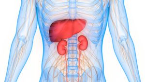
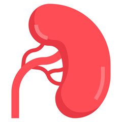
Abdominal ultrasound exams are a valuable diagnostic tool for scanning the kidneys. An abdominal ultrasound exam can be an important step in the diagnostic process for patients who may be experiencing kidney-related issues or abnormalities. The imaging and observation of the kidneys through an abdominal ultrasound exam is a reliable diagnostic tool, as it can detect many pathologies in the area.

With the use of an abdominal ultrasound exam, physicians can observe any abnormalities in the kidneys such as cysts, masses, stones, or other issues that may be affecting their overall function. This diagnostic tool is a non-invasive way to examine the kidneys, with no radiation or injections of chemicals required, making it a safe option for patients. For a Kidney (Renal) examination, the patient must present with a full bladder. The bladder is a part of the renal system and as practitioners, we must assess that it is functioning properly.
Upon arrival, the patients bladder can be scanned first. The overall volume can be recorded and bilateral urethral jets should be noted with doppler. If there is no pathology detected, the patient is free to empty his/her bladder fully. We want to ensure the patient is voiding properly. If there is any fluid retained after voiding, a volume should be recorded.
For the patients Kidney exam: Both Kidneys will again be scanned in Transverse and Longitudinal planes. For the average adult a 2-6MHz curved array transducer will be required. For pediatric patients, a linear array transducer with a higher frequency will allow for shallower scanning. The position of the patients will depend on a combination of things. a) patient body habitus b) what position delivers to you the best view based on the patient’s anatomy. c) How well the kidneys are visualized based on respiration instructions given to your patients. “take a deep breath in…. and out”… This will allow you to exam the kidney as it changes positions with changes in the diaphragm.
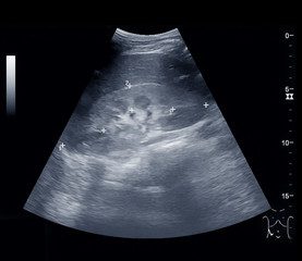
The right kidney is usually found more caudally and is generally more slim to the left kidney. The kidney is divided into parenchyma and renal sinus. The sinus is hyperechoic and composed of calyces, renal pelvis, fat, and intrarenal vessels. Generally you should not be able to visualize the urinary collecting system within the renal sinus. If it is abnormal, due to a blockage, it will be filled with fluid, thus being quite visible to the eye.
The parenchyma is more hypoechoic and homogeneous. It is divided into an outer cortex and innermost and slightly less echogenic medullary pyramids. cortex. To separate the pyramids, are corticals called columns of Bertin. A normal length of an adult kidney is generally 10-13cm. For children, . The cortex should be estimated from the base of the pyramid and should be between 7-10mm.
Doppler of the kidneys is widely used.This evaluates perfusion. Colour doppler along with spectral
 doppler is of common practice. Spectral doppler should be applied to the renal artery and selected interlobular arteries. Peak systolic velocities, resistive index, and acceleration curves can be estimated . Peak systolic velocity of the renal artey above 180cm/s is a predictor of renal artery stenosis of more than 60%. The resistive index above 0.70 is indicative of abnormal renovascular resistance.
Renal pathologies: fifty percent of people over fifty have renal cysts. They are benign fluid filled sacs that do not require any further evaluation. If they are complex cysts, meaning they have membranes dividing the fluid-filled centre, calcification ,or irregular thickened walls, they may be followed up. We would also use doppler. If a patient presents with a solid mass, generally they are followed up with a contrast-enhanced CT and referred to the radiologist for further evaluation.
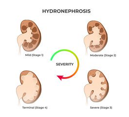
Hydronephrosis is the enlargement of the urinary collecting system usually related to urinary obstruction and can include the pelvis, calyces, and the ureters. It can be caused by anything compressing on the ureter, such as a mass, abscess, or tumour. Another cause can be Renal Calculi. Ultrasound has a lower sensitivity and specificity for detecting renal stones than CT, but it is recommended that it be used as a first attempt due to it’s no risk to patients in radiation exposure. When there are larger stones (5-7mm and larger) accompanied by hydronephrosis, the accuracy becomes higher.
Chronic Kidney Diseases, Acute Renal Injury, and Ultrasound Guided procedures are also quite common exams done with the use of ultrasound. We are able to asses the biopsies, abscess drainages, and oversea many other procedures. Dialysis patients are scanned frequently to assess the function of their kidneys. Trauma of any organ is quickly assessed with the use of ultrasound imaging. We are able to analyze to some extent the damage that has occurred to an organ within moments. Ultrasound has truly become a force to be reckoned with.
Scanning the Pancreas

The pancreas is also part of a routine abdominal ultrasound exam. Sonographers have the capability to perform upper abdominal ultrasound examinations accurately, which can be used as a diagnostic tool in General Practice. Abdominal ultrasound provides a safe, non-invasive way to visualize and diagnose the pancreas. During the exam, the sonographer will use high-frequency sound waves to create an image of the pancreas. This image will allow for the detection of any abnormalities or irregularities in this organ. Unfortunately, for a lot of patients, this organ is difficult to visualize. It is located in a region where there is consistently a lot of bowel gas present exactly where the pancreas lives and needs to be imaged. For this reason, we have the patient fast, in hopes to reduce this interference from overlying bowel gases.
We use a 3-6 MHz curvilinear transducer. The pancreas consists of a head, body, and tail. For the Pancreatic body, we can attempt to use the left lobe of the liver as an acoustic window. The patient is asked to take a deep inspiration and/or push their “belly out, as to make a beer belly.” This can sometimes disperse the gas away from the image. For the head of the pancreas, a right subcostal approach with the transducer angled medially can sometimes help. If the patient has a dilated common bile duct, you will often see it running through the head of the pancreas into the body. The pancreatic tail can be extremely difficult to visualize. Sometimes, a water filled stomach can be used as a window. Try to scan coronally, in a right lateral decubitus position using the spleen as an acoustic window.
Measurements: head=34mm, body=29mm, tail=32mm, length=12-20mm, pancreatic duct=less than or equal to 3mm. The pancreas become a more echogenic with age due to fatty replacement.
Pathologies: pancreatitis, masses, cysts, calcifications, enlargement, cancer, anything abnormal in appearance.
Note: the key is to know what normal looks like. Then you know when something is not supposed to be there. Document it, Report it, and get the patient where they need to be to get the help they need.
Scanning The Gallbladder and Common Bile Duct

When it comes to acing an abdominal ultrasound exam, it’s important to have a strong understanding of the anatomy of the gallbladder and common bile duct. Before beginning the exam, it’s essential to familiarize yourself with the location and function of these organs to ensure accurate imaging.
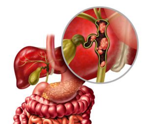
The gallbladder stores and concentrates bile from the liver. The bile is then released into the first section of the small intestine (the duodenum), where it helps your body to break down and absorb fats from food. A tube that carries bile from the liver and gallbladder, through the pancreas, and into the small intestine. The bile ducts carry bile from your liver to your small intestine. When bile ducts become damaged, bile can back up into the liver, causing damage to liver cells. This damage can lead to liver failure. CBD should be less than 7mm AP.
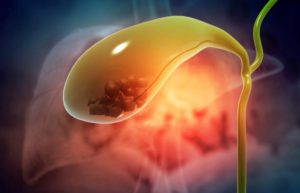
During the exam, it’s important to use proper scanning techniques in both the transverse and longitudinal planes to capture a complete image. Additionally, it’s crucial to pay attention to any abnormalities in the size, shape, echogenicity, or wall thickness of the gallbladder. Using colour Doppler imaging can also help identify any stones, polyps, masses, or sludge present within the gallbladder. It should be documented whether or not any of the echogenicities are mobile or not by having the patient go from a prone to standing position while scanning.
Measure any dilated structures and note their location relative to other organs. A precise and thorough exam will ensure optimal image quality and help identify any potential issues. With proper preparation and attention to detail, sonographers can successfully ace abdominal ultrasound exams every time.
Scanning Bowel and Displacing Bowel Gas While Scanning
One of the challenges of abdominal ultrasound is ensuring clear imaging by avoiding gas in the bowel. To prevent this, it is recommended to have the patient take a deep breath and hold it during the imaging process. If gas does not clear, the patient may need to be placed in a different position to ensure clearer imaging. The Valsalva maneuver is also used commonly as a technique to displace gas. It is essentially having the patient bare down on forced expiration…(kind of like when attempting to clear your ears after a long flight.)
By attempting these different tricks and properly scanning bowel and avoiding gas, sonographers can increase their chances of achieving a productive abdominal ultrasound exam.
Difficulties faces with patients of different body habitus
Conducting an abdominal ultrasound needs a lot of care and process. The person conducting the test must have knowledge about the patients’ body types to deliver accurate and proper images. An obese patient’s excess tissue can create air pockets, obstructing the quality of the ultrasound images. Conversely, thin patients may make it challenging to access organs due to insufficient tissue. An elderly patient’s abdominal anatomy can bring challenges in identifying an area of interest, such as atrophied muscles, which may be hard to see. Children tend to move during the procedure, which results in blurred photos or misdiagnosis. Despite these difficulties, patients should always undergo abdominal ultrasounds from certified sonographers as the images can help identify and treat underlying health issues.
What Ultrasound transducer to select and frequency to use
Selecting the appropriate ultrasound transducer and frequency for abdominal ultrasound exams is crucial to obtain high-quality images. High-frequency transducers are recommended to improve image quality. General practitioners should use abdominal ultrasound as a diagnostic tool to identify potential stomach and liver problems. Sonographers should have an understanding of sonographic principles and applications when selecting the transducer. A prospective observation of the patient’s abdominal area should be done to determine the best approach and frequency to use. Furthermore, tissue harmonic imaging sonography should be considered when deciding on the type of transducer to employ for abdominal ultrasound exams. Overall, using the right transducer and frequency during abdominal ultrasound exams can help sonographers to accurately diagnose their patients.
Common pathologies seen on abdominal ultrasound exams
An abdominal ultrasound is a non-invasive procedure that uses high-frequency sound waves to produce images of the internal organs in real-time. The procedure can detect a variety of pathologies, and some of the common pathologies related to an abdominal ultrasound exam include renal stones, cystic lesions, and cirrhosis. The exam is also useful for diagnosing aortic aneurysms, measuring the size of the spleen, and identifying fluid collections. In addition to its diagnostic abilities, abdominal ultrasound is helpful in guiding interventional procedures, such as biopsies or drainages. Emergency departments take advantage of the ultrasound’s quick response to suspected pathologies to provide faster and accurate diagnosis, resulting in enhanced treatment planning. To sum up, with its excellent diagnostic and interventional capabilities, abdominal ultrasound is an essential tool for sonographers and other medical professionals in identifying health complications in the abdominal area.
Common reasons for performing abdominal ultrasound exams
An abdominal ultrasound is a common diagnostic imaging procedure that helps doctors detect and diagnose various abdominal issues. Ultrasound is preferred as it is radiation-free and provides images in real-time. Abdominal ultrasounds are commonly used to diagnose abdominal pain, masses, and to assess organ function, such as the liver or gallbladder. They can also detect abnormalities in the kidneys, bladder, or other organs. In some cases, ultrasounds may help diagnose certain types of cancers like ovarian, pancreatic, or liver cancers. Additionally, they are used to detect blood clots, aneurysms, or other blockages in the abdominal area. An abdominal ultrasound is a non-invasive procedure making it safe, quick, and easy to perform. Regular checkups with your physician can help you stay on top of any potential health concerns.
Frequently Asked Questions
How do you choose a good quality abdominal ultrasound machine and accessories?
Choosing a good quality abdominal ultrasound machine and accessories can be a difficult task. There are several factors to consider before making a purchase, such as the intended use, image quality, portability, user-friendliness, and cost. Here are some tips to help you choose a good quality abdominal ultrasound machine and accessories:
1. Determine your needs: Consider the primary use of the ultrasound machine and the specific features you need. For abdominal ultrasounds, you may want a machine that offers clear imaging and ease of use.
2. Look at image quality: The image quality of an ultrasound machine is important to get an accurate and clear diagnosis. Check the pixel count, resolution, and image depth.
3. Consider portability: Depending on your needs, you may want an ultrasound machine that is portable and easy to move to different locations.
4. Check user-friendliness: The system should be user-friendly with an easy-to-use interface.
5. Compare with other options: Look at different models and compare them based on the parameters that are most important to you.
6. Check reviews: Online reviews from other customers or users can provide valuable insight into the quality and performance of the machine.
7. Look at the warranty and after-sales service: A product with a good warranty and after-sales service can give you peace of mind and assist you in case of any issues.
What is the protocol for abdominal ultrasound?
The protocol for abdominal ultrasound may vary depending on the reason for the exam, but generally includes the following steps:
1. The patient will lie on their back or side on an exam table with their abdomen exposed.
2. A gel will be applied to the skin over the abdomen to help transmit sound waves from the ultrasound transducer.
3. The sonographer will move the transducer over the abdomen while investigating all organs, taking images of various organs and structures as they go.
4. The images will be displayed on a monitor for the sonographer to view and analyze.
5. The exam may take anywhere from 30 minutes to an hour, depending on the complexity of the scan and the condition being investigated. The preliminary report and images are then given to a Radiologist for final reporting.
It is important to follow any pre-exam instructions provided by your healthcare provider, such as fasting or drinking water before the exam. After the exam, you may be asked to wait for a short time to ensure that all necessary images have been captured. Your healthcare provider will then review the images and discuss the results with you.
How do you study for an abdominal ultrasound?
If you are studying for an abdominal ultrasound, there are several things you can do to prepare:
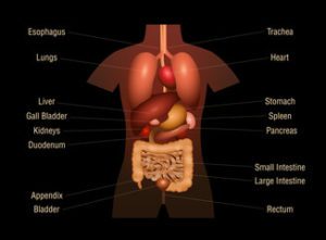
1. Review the anatomy of the abdomen: Understanding the structures and organs in the abdomen, such as the liver, pancreas, and gallbladder, will help you identify them on the ultrasound images.
2. Familiarize yourself with ultrasound terminology: There are specific terms and abbreviations used in ultrasound reports, so it’s important to be comfortable with them.
3. Practice scanning techniques: If you have access to an ultrasound machine, practice scanning techniques to improve your ability to obtain high-quality images.
4. Study sample images: Look at sample images and compare them to corresponding anatomy diagrams to reinforce your understanding of what you are seeing.
5. Enroll in a training program: If you are new to ultrasound or want to reinforce your skills, taking a training course can be helpful.
Remember, ultrasound technology is constantly evolving, and new techniques and equipment are being developed all the time. Staying up-to-date on the latest advances in ultrasound technology can help you provide the best possible care for your patients.
Where can I find a cheat sheet on performing an abdominal ultrasound for sonographer?
There are a number of resources available online that can provide a helpful cheat sheet for performing an abdominal ultrasound as a sonographer. Some of the best places to look include online ultrasound training programs, professional associations for sonographers, and medical imaging forums and websites. You might also consider reaching out to colleagues in the field or consulting with a mentor who can provide guidance and advice on best practices for performing abdominal ultrasounds. Ultimately, the key to success as a sonographer is to continue learning and staying up-to-date on the latest techniques and technologies in the field. You can also feel free to connect with me, as I have a lot of years of experience in the field.
What are the benefits of performing abdominal ultrasound for sonographer?
Abdominal ultrasound is a common diagnostic tool used by sonographers to visualize the abdominal organs and identify any abnormalities or medical conditions. Some of the benefits of performing abdominal ultrasound for sonographers include:
1. Non-invasive: Unlike other diagnostic imaging tests, such as CT scans or MRI scans, abdominal ultrasound is non-invasive and does not use ionizing radiation, making it a safer and less invasive option for patients.
2. Real-time imaging: Abdominal ultrasound provides real-time images of the abdominal organs, allowing sonographers to observe the movement and function of the organs in real-time.
3. Cost-effective: Abdominal ultrasound is generally less expensive than other diagnostic imaging tests, making it a cost-effective option for both patients and healthcare providers.
4. Early detection of medical conditions: Abdominal ultrasound can help detect medical conditions early on, when they are most treatable, providing a better prognosis for patients.
5. Portable: Abdominal ultrasound equipment is portable and can be used in a variety of settings, making it a convenient option for providing diagnostic imaging services in remote or underserved areas.
What are some common abnormalities or conditions that can be detected through an abdominal ultrasound?
Abdominal ultrasounds can help detect various abnormalities and medical conditions, including gallstones, liver disease, pancreatic cancer, abdominal aortic aneurysm, and kidney stones. They can also be used to evaluate the size and condition of organs such as the liver, pancreas, spleen, and kidneys, and to detect fluid or swelling in the abdomen. Overall, abdominal ultrasounds are a useful diagnostic tool that can help healthcare professionals identify potential health issues early on.
What is the role of the patient during an abdominal ultrasound exam?
During an abdominal ultrasound exam, the role of the patient is to lie still on the exam table while the healthcare provider moves the ultrasound wand over the abdomen to capture images of the internal organs. The patient may be asked to change positions or hold their breath at times to get better images. It is important to follow any instructions given by the healthcare provider and let them know if you experience any discomfort during the exam.
Conclusion
In order to ace your abdominal ultrasound exam, proper preparation is key. From scanning the liver to avoiding gas in the bowel, there are many different techniques and skills you need to master. Additionally, understanding common pathologies and why an abdominal ultrasound exam may be necessary is crucial. And no matter what the patient’s body habitus, it is important to select the right ultrasound transducer and frequency for the best imaging results. With a little bit of practice, you can be sure to excel at your next abdominal ultrasound exam.

